
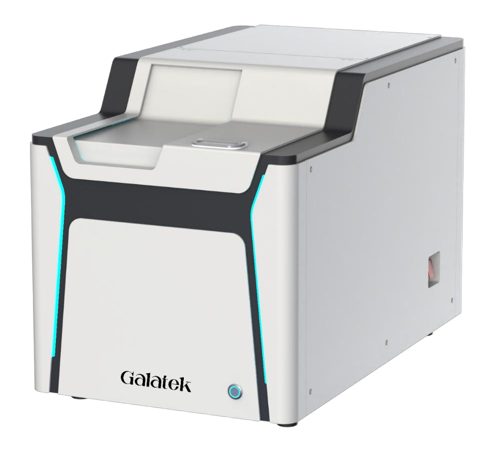
G-Cell T3000 Confocal
T3000 uses spinning-disk confocal microscopy, five lasers (405, 488, 561, 638 and 730 nm) and an sCMOS camera.
3D imaging and AI analysis
G-Cell combines 3D confocal imaging of cells and organoids, time-lapse live-cell observation and AI-assisted image analysis. A large field of view reduces the number of images required for scanning.
Scope of work
T3000 uses spinning-disk confocal microscopy, five lasers (405, 488, 561, 638 and 730 nm) and an sCMOS camera.
T2000 uses white light for brightfield imaging, five LED channels and a CMOS camera; an optional laser source is available for 561 nm.
Laser and image-based autofocus sets focus for different samples and vessels, reducing manual adjustments.
The software enables 3D reconstruction, object recognition and image segmentation. More than 60 processing and analysis modules are used to measure morphology, signal intensity and texture. AI models can also be trained for specific sample types.
Adapt the system to your samples: choose widefield (2D) or confocal (3D) mode and add an incubation chamber if you conduct long-term live-cell observations.
Swipe the table sideways to see all columns.
| Feature | T2000 Widefield | T3000 Confocal |
|---|---|---|
| Imaging mode | Widefield | Confocal and widefield — switchable |
| Fluorescence illumination | Five LED channels; optional 561 nm laser | Five lasers: 405, 488, 561, 638 and 730 nm |
| Camera | CMOS | sCMOS |
| Objectives | Automated four-position objective turret | Automated four-position objective turret |
| Live-cell observation | Optional temperature, humidity and CO₂ control module | Optional temperature, humidity and CO₂ control module |
Technical documentation
Contact
Describe your process and the scope of planned automation.